A choroid plexus cyst is a small fluid filled space that occurs in a gland in the brain called the choroid plexus. In response to increased bone resorption due to excess parathyroid hormone pth secretion and.
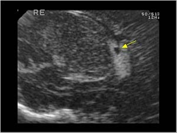
 Choroid Plexus Cyst Radiology Case Radiopaedia Org
It uses the datamuse api to find related words and then finds combinations of these words that pair well together phonetically.
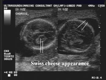
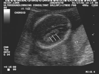
Choroid plexus cyst ultrasound picture. Port manteaux was created by sean gerrish and doug beeferman. Cri du chat syndrome cdcs is a genetic disease resulting from a deletion of the short arm of chromosome 5 5p. The rostrum is the last part.
The common cyst that forms from a hair follicle. In 1963 the most important clinical features are a high pitched cat like cry hence the name of the syndrome distinct facial dysmorphism microcephaly and severe psychomotor and mental. Dysgenesis which may be complete or partial is a result of encephalomalacia secondary to toxic ischemic or traumatic events 2.
This term and pattern are distinctive for hyperparathyroidism. Its clinical and cytogenetic aspects were first described by lejeune et al. Rugger jersey spine describes the prominent endplate densities at multiple contiguous vertebral levels to produce an alternating sclerotic lucent sclerotic appearance.
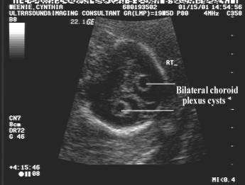
Small pinched off blebs that are formed when the brain is developing the choroid plexus. Summary recurrent bacterial meningitis is a rare phenomenon and generally poses a considerable diagnostic challenge to the clinician. Choroid plexus cysts cpc a fluid filled space in the brain which appears as a cyst and is not considered a brain abnormality which is more commonly associated with trisomy 18 renal pyelectasis the main area of a kidney is enlarged.
Ultimately a structured approach and early diagnosis of any underlying pathology are crucial to prevent further episodes and improve the overall outcome for the affected individual. This gland is located on both the left and right side of the brain and it makes. A cyst in the lumbar spine that may cause symptoms of spinal stenosis.
They contain cerebrospinal fluid. The impact factor 2018 of scientific reports is 4 011 which is just updated in 2019 compared with historical impact factor the impact factor 2018 of scientific reports dropped by 2 69 the impact factor quartile of scientific reports is q1 the impact factor if or journal impact factor jif of an academic journal is a scientometric index that reflects the yearly average number of. It begins with the genu and then continues posteriorly along the body to the splenium.
The development of the corpus callosum occurs between the 12 th and 16 20 th weeks of gestation 2 4. Dandy and blackfan demonstrated the csf production within the ventricles by choroid plexus and divided hydrocephalus into communicating and non communicating types 5. The algorithm tries reconstruct a spelling for the new word after generating its pronunciation and sometimes this spelling isn t quite right.
In this article we are reviewing the existing literature on this topic over. This mimics the horizontal stripes of a rugby jersey. This classification was made based upon the appearance of phenolsulfonphthein psp in the lumbar csf space after introducing it in the ventricle.
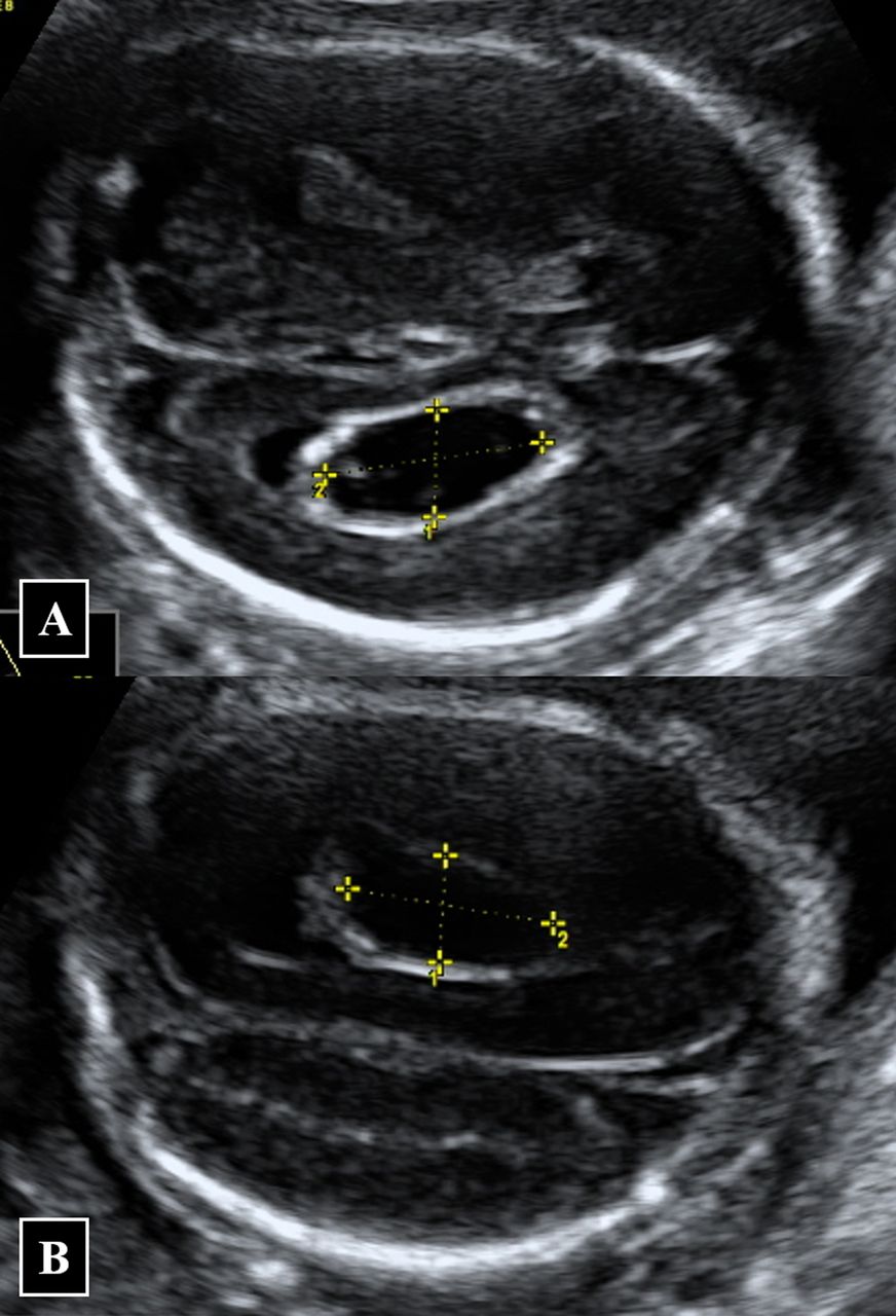
 Choroid Plexus Cyst Antenatal Radiology Reference Article
 Choroid Plexus Cyst Antenatal Radiology Reference Article
Pediatrics 9 6 Neonatal Brain And Spine Case 9 6 7
 Choroid Plexus Cyst Youtube
 Choroid Plexus Cyst Looking Through A Transducer
 Isolated Fetal Choroid Plexus Cysts Contemporary Obgyn
 Andrea Arch Ultrasound Finding Choroid Plexus Cyst
 Wk 6 L 2 Choroid Plexus Cyst Radiology Case Radiopaedia Org
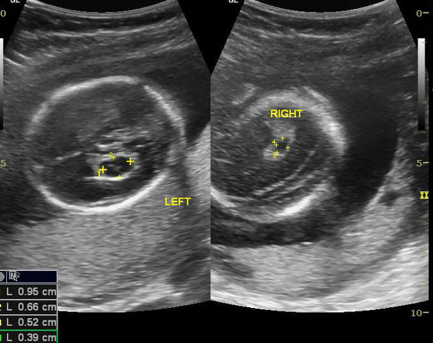
 Isolated Large Bilateral Choroid Plexus Cysts Associated With
 Choroid Plexus Cyst Radiology Case Radiopaedia Org
 Scary Pregnancy Vlog Weeks 14 20 Ultrasound Choroid Plexus Cyst
 Global Library Of Women S Medicine
 Ultrasonographic Soft Markers Of Aneuploidy In Second Trimester
 Medpix Case Trisomy 18
 Ultrasound Images Of Fetal Brain
 Chromosomal Anomalies Radiology Key